microscope root canal treatment
Root canal is a ‘life saving’ procedure for severely decayed or damaged teeth. It is one of the most common dental procedures we perform in our clinic and this process can save your natural tooth and prevent the need for dental implants or bridges. As root canal specialists (endodontists) Dr. Vishwas and Dr. Shivani always use the latest techniques and materials including performing the procedure under a microscope (Carl Zeiss OPMI pico) to ensure a smooth root canal therapy which many people report is no more uncomfortable than getting a dental filling.
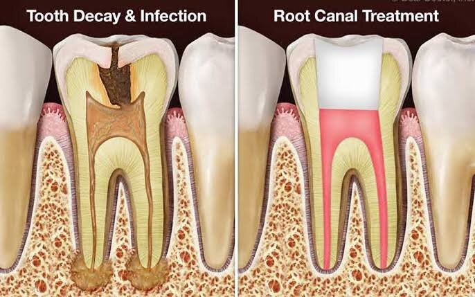
1. What is root canal treatment?
- Endodontic/ root canal therapy (RCT) is a procedure that is performed by a root canal specialist (endodontist) in cases where the nerve of the tooth is affected and the only way to save the tooth is by removing the affected nerve and replacing it with a filling. Your tooth is made up of three primary layers- the outer hard white enamel, the middle yellow dentine and the innermost nerve called the pulp. The pulp is essentially a collection of blood vessels that help to build the surrounding tooth. Infection to the pulp can be caused by deep decay, trauma, cracks/tooth fracture, old amalgam fillings and even repeated dental procedures. This can result in visible injury or swelling of the tooth, pain, sensitivity to water or temperature, gum pain, food lodgement and sometimes even no symptoms at all. In some situations you may have transient symptoms which when ignored results in progression of the infection which finally results in such severe damage to the tooth that RCT will be your only option to save the tooth.
Once a diagnosis has been made and a root canal treatment is advised- our root canal specialist (endodontist) will first completely numb the tooth to be treated. The decay is removed using specialized tools and the tooth is completely cleaned using a combination of aseptic techniques and irrigating solutions under rubber dam (for isolation). The tooth is then filled with a biocompatible material and is then ready to receive a crown. The entire process is performed under a microscope since microscope root canal treatment has been proven to be more precise, effective and free of any operator errors. A root canal treated tooth should always be three dimensionally sealed and protected with a crown (cap) so as to prevent reinfection and so that you can resume normal eating on the tooth.
2. Should I save my tooth?
- Root canal therapy is an attempt to save a damaged tooth which would otherwise be extracted. If a tooth is removed, the replacement which would be an implant or bridge has a success and failure rate, with their own set of challenges. Also placing an implant or bridge will require significantly more time and may require additional procedures to neighboring teeth and supporting tissues. No replacement, no matter how well fabricated, is as good as your natural tooth. Should a reasonable success rate be expected with the microscope root canal, it is always best to try and save your tooth. The alternatives will still be available if the tooth does not respond to the root canal but not the other way around. It is always better to save the tooth with RCT since modern endodontic techniques have made the procedure virtually painless and comfortable. Visit Summit Dental Clinic to learn more about microscope root canal procedure.
3. Will root canal treatment hurt?
- No. Root canal treatment does not cause pain, it relieves it. People are often worried about the prospects of pain during root canal therapy and the key to a painless procedure is definitive local anesthesia. Dr. Vishwas and Dr. Shivani always use a combination of numbing gel and local anesthetic agent to completely numb the area with the latest techniques and the anesthesia will be confirmed before beginning the procedure. They strongly believe that that painless root canal treatment / endodontic therapy is possible with today’s technology and with the understanding of the anatomy and physiology of the oral neurorovasculature.
4. How long will the procedure take?
- Although each tooth is unique and the time required to complete the treatment is dependent on the level of decay in the tooth, you should plan on spending about an hour and a half at our office during your root canal treatment appointment. Most microscope root canal procedures are completed in one sitting, unless there is severe infection, procedural difficulties or in cases of re-root canal treatments. At Summit Dental, we understand that your time is valuable and we will do our best to render treatment as expeditiously as possible without compromising on the quality of your care. We also try our best to minimize your waiting time at our clinic and we appreciate your understanding and patience when we try to do everything we can to accommodate you and your needs.
5. As a patient, what should I expect from Summit Dental Clinic?
- You should expect to be treated with courtesy and respect. You should feel comfortable all the time with our dental team and ability to handle your care professionally while being personable and attentive. You have the right to a painless procedure and personal attention by Dr. Shivani and Dr. Vishwas as well as their staff. Depending on the kind of infection inside the tooth, there be be anywhere from mild to moderate discomfort after the microscope root canal procedure or in-between visits. This is due to the healing process at the end of the root. We give you precise post-operative instructions to minimize this discomfort.
6. What happens after a microscope root canal procedure?
- When your root canal therapy has been completed, you will experience some discomfort for 2-3 days following the procedure- this is normal and is just the healing process of the tooth. This discomfort can be minimized with painkillers which will give you immediate relief. You can drive back and resume work following your procedure. The next step will be shaping the tooth and taking an impression or scan to receive a crown. Our dental team will explain the different crowns available, the benefits and disadvantages of each material and the price range for each type of crown. Failure to properly restore and protect the tooth with a crown or filling may result in re-infection or fracture of the treated tooth. Once a crown has been cemented or bonded, you can resume normal chewing, brushing and flossing on the root canal treated tooth. It is rare for people to experience complications after routine endodontic treatment but can occur if you do not follow our precise instructions. To prevent further decay, continue to practice good dental hygiene.
7. I am worried about X-rays. Should I be?
- No. While x-rays are necessary during your microscope root canal treatment. We use advanced digital radiographic systems which minimizes the radiation levels up to 80-90% lower than that of conventional dental X-ray machinery (approximately 9 digital radiographs equals 1 film radiograph!). These digital images can be e-mailed to you so that you can keep a record of the treatment done as well as a reference to compare the healing of the tooth and bone. We are extremely particular about patient and doctor safety in our dental clinic and provide you with a lead apron as well as thyroid collar while taking the X-rays. Please inform our dental staff at the time of the consultation if you are pregnant.
8. What new technologies are being used?

- Dr. Vishwas and Dr. Shivani are highly trained and skilled in using the latest technology to deliver the best standard of care. They are constantly updating themselves with the latest from the field of endodontics and cosmetic dentistry so that they can incorporate it at Summit Dental to make your treatment as comfortable and transparent as possible.
Operating microscope- In addition to digital radiographs which reduce the x-ray exposure, we also utilize operating microscopes (Carl Zeiss OPMI pico) which magnifies our field of view up to 30 times and illuminates our field with Xenon fiber optic illumination. This combination allows for exceptional diagnostic technical ability to treat the tiny root canal spaces inside your tooth. Such microsurgical techniques have revolutionized endodontic care within the last ten years and allows for a far superior quality of care today than ever before. Today, problems such as cracks, root fractures and calcified canals can be better visualized and therefore treated. Also a video camera on the operating microscope can record images of your tooth to further document our findings. It is our belief that RCT should only be performed under these microscopes for the highest quality of care to be rendered.
Apex locators- these machines allow for the electronic and accurate measurement of the root length to precisely verify the radiographic findings. They improve the quality of care and allow for more precision based endodontics.
Sonic activation- electronic instruments are now available which create sonic vibrations, which clean the root canals more effectively and efficiently. These instruments increase the quality of care and allow for success in situations that were previously hopeless.
Nickle Titanium instrumentation- Today, the cleaning and shaping of root canals is no longer a brute procedure done with hand files. The job is now down with mechanized, engine driven rotary files that have made the procedure more efficient, predictable, quieter and far more comfortable than ever before.